
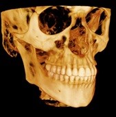
Conebeam CT scanners deliver very accurate Cranio-Maxillofacial images and combines “the power of focused-field 3D with cutting-edge panoramic imaging” for enhanced diagnostic capabilities. Our conebeam CT scanner can capture both jaws (9cm x 15cm), or one of the largest fields of vision, the complete dento-maxillofacial (18cm x 20cm) complex. This allows for a precise evaluation of the facial bones and the soft tissues associated with them. Therefore, implant placement is more precise, facial bone fractures can be diagnosed, and pathology can be evaluated.
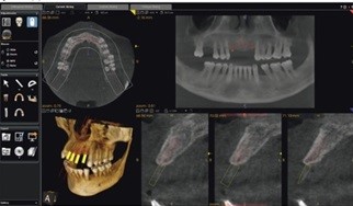
This system allows capture of clear, detailed, anatomically correct 3D images in seconds. Comprehensive imaging software then enables rapid onscreen access of both 2D and 3D images; we can select the desired program, and the unit automatically uses the correct sensor.
The high-resolution images for each tooth let the clinician view even the smallest details from all angles and slices, while extended field-of-view capabilities enable capture of the entire mandibular or maxillary arch. This increases the level of detailed clinical information available to us, while mitigating the level of patient radiation exposure.
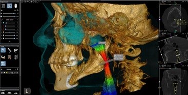
Our conebeam CT scan can also perform airway analysis for patients with sleep apnea, by providing a colorful three-dimensional view of your upper airway. Our airway analysis automatically segments and measures the upper airway. Automatically calculates total volume, minimal cross-sectional area, anterior/posterior and left/right measurements. Displays and updates measurement values in real time while browsing slices or changing angulations.
Contact our office for more information today.
Damone E. Smith, D.D.S
Diplomate, American Board of Oral and Maxillofacial Surgery
Adjunct Professor of Oral and Maxillofacial Surgery
Nova Southeastern University School of Dentistry
954-838-0621
Florida Center for Oral Surgery & Dental Implants